This model extended so the aim of the present study was to design of knee joint model to cover educational aspect so that student radiographers can easily imagine the different posture during imaging of knee joint. The kneecap the patella joins the femur to form a third joint called the patellofemoral joint.
Solved Review Sheet 11 7 Label The Photograph Of A Knee Chegg Com
The kneecap patella forms the protective bony covering for the knee joint.
. Notch located inferior to the ischial spine 01 t where the patellar ligament attaches I 12 kneecap 13. The knee is a complex joint that flexes extends and twists slightly from side to side. Anatomy and Physiology questions and answers.
3B Scientific Functional Models of the shoulder elbow hip and knee have flexible ligaments and provide a graphic demonstration of the anatomy and mechanics of the major joints allowing better doctor-patient or teacher-student understanding. The shoulder joint is built for mobility. The knee joint has three parts.
This is an online quiz called Knee Joint Model Labeling Quiz. Tibial collateral ligament 8. Considering the structure of the menisci would you expect.
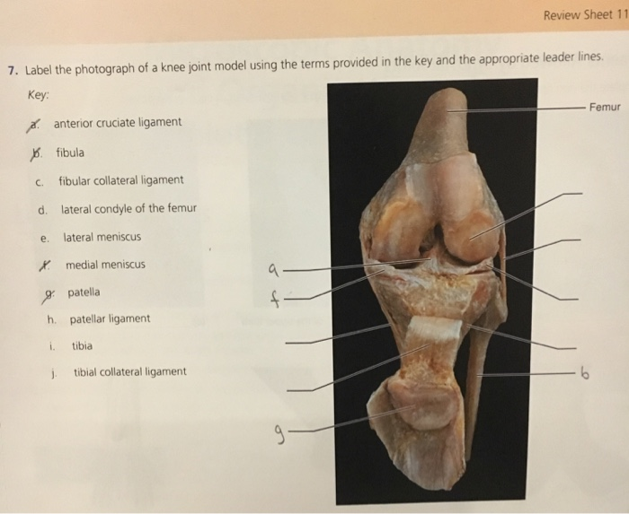
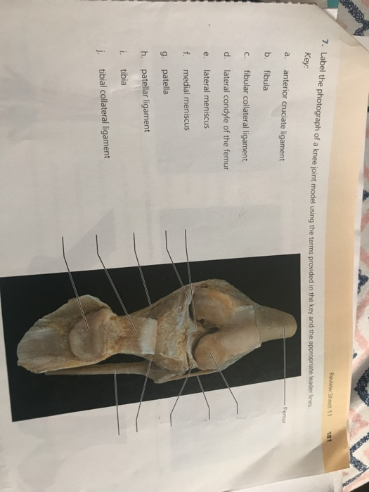
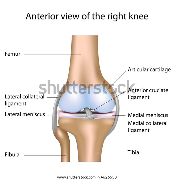
The upper leg bone femur and the larger lower leg bone tibia are connected via the knee joint. 181 Review Sheet 11 ed in the key and the appropriate leader nes anterior cruciate ligament fibula fibular collateral ligament lateral condyle of the femur lateral meniscus medial meniscus patella patellar ligament i tibia j. The color of the natural-cast bones of the knee joint is extremely realistic.
1000 960 pixels 33 32 in DPI 300 JPG. Sacroiliac joint talus tarsals tibia tibial tuberosity of the thigh bone 6. The knee joint is the hinged joint that allows the legs to bend.
Whether you choose life-size or half-size these fully flexible joint models demonstrate abduction ante-version retro-version. Longest strongest bone in body 8. Female Pelvic Skeleton with Movable Femur Heads - 3B Smart Anatomy.
Knee joint anatomy labeled. Deluxe Functional Knee Joint Model. Shoulder Joint With Rotator Cuff 5-Part.
This model consists of portions of the femur tibia and fibula menisci and patella. All these parts combine and work together. The cartilage on the knee joint surfaces is marked blue.
The knee is the meeting point of the femur thigh bone in the upper leg and the tibia shinbone in. Subscribe Today and Get Industry-Leading Content Support and Licensing. This model clearly demonstrates abductions anteversion retroversion internal and external rotation.
Fibular collateral ligament d. The femur tibia and patella. Label the photograph of a knee joint model using the terms provided Key.
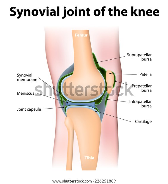
A fibrous capsule surrounds the knee joint and a series of ligaments holds the. There is a printable worksheet available for download here so you can take the quiz with pen and paper. Learn vocabulary terms and more with flashcards games and other study tools.
500 480 pixels 17 16 in DPI 300 JPG. D The menisci in the knee joint c an be torn for a variety of reasons. Ad Boost Your Brands Digital Presence Now Get Authentic Local Images for Targeted Results.
Because the knee supports almost all of ones body weight this joint is highly susceptible to injury. It is a complex hinge joint composed of two articulations. Thin lateral leg bone permits passage of the sciatic nerve 12 SSer 10.
Fibula c fibular collateral ligament d lateral condyle of the femur e. Find Knee Joint Anatomy Labeled stock images in HD and millions of other royalty-free. MSRP 11000 MSRP 11000 10100.
Click on a photo for a larger view of the model. With the help of knee models however it is easy to see how the three main components the femur patella and tibia work together and how they can buckle under stressful situations. This knee joint model also clearly defines the ACL and PCL.
Click on L abel for the labeled model. Back to Muscular System. The thigh bone the femur meets the large shin bone the tibia to form the main knee joint.
Patellar ligament i tibia j. The tibiofemoral joint is an articulation between the tibia and the femur while the patellofemoral joint is an articulation between the patella. The knee also known as the tibiofemoral joint is a synovial hinge joint formed between three bones.
The tibiofemoral joint and patellofemoral joint. MSRP 16100 MSRP 16100 14800. Your Skills Rank.
Damage in even one part can hinder the functioning of the knee. Anterior cruciate ligament b. MSRP 32400 MSRP 32400 29600.
7 Knee joint modeling using OpenSim software 71 Overview of OpenSim software 7-2 72 Development of a skeletal model of the knee joint 7-3 73 Inverse kinematic analysis 7-5 74 Muscle modeling 7-7 75 Contact forces modeling and analysis 7-11 76 Summary and discussion 7-20 References 7-22 Several computational codes such as SIMPACK Rulka 1990 AnyBody. This is a free printable worksheet in PDF format and holds a printable version of the quiz Knee Joint Model Labeling QuizBy printing out this quiz and taking it with pen and paper creates for a good variation to only playing it online. Label the diagram of a typical synovial joint using the terms provided in the key and the appropriate leader lines.
The anatomy of the knee consists of bones muscles nerves cartilages tendons and ligaments. Materials and Methods The basic element of the model structure was gypsum in its powder form as well as water for mixing and as a. Movements at the knee joint are essential to many everyday activities including walking running sitting and standing.
This joint has an inner medial and an outer lateral compartment. Joint between axial skeleton and pelvic girdle 7. Knee joint is one of the most important hinge joints of our body.
Anterior cruciate ligament b. Functional Knee Joint Model. Label the photograph of a knee joint model using the terms provided in the key and the appropriate leader lines Key Femur a.
Picture of Knee Joint. Label the photograph of a knee joint model using the terms provided in the key and the appropriate leader lines. Two rounded convex processes known as condyles on the distal end of the femur meet two.
Help you to identify the location of internal structures and understand the relationship between and among the organs. Knee education models are useful classroom. The femur tibia and patella.
Lateral condyle of the femur Lt side Rt side - A - - F - - - - - - -. 5001 4800 pixels 167 16 in DPI 300 JPG. Start studying Knee Joint Label.
The knee joint is a synovial joint that connects three bones. A person with a high degree of muscular strength generally also has a high degree of muscular endurance. Its complexity and its efficiency is the best example of Gods creation.
Picture of Knee Joint.
Solved 7 Label The Photograph Of A Knee Joint Model Using Chegg Com

Knee Joint Label Pictures Flashcards Quizlet
Knee Joint Anatomy Labeled Stock Illustration 157672166 Shutterstock
Knee Joint Anatomy Labeled Stock Illustration 157672166 Shutterstock


0 Comments